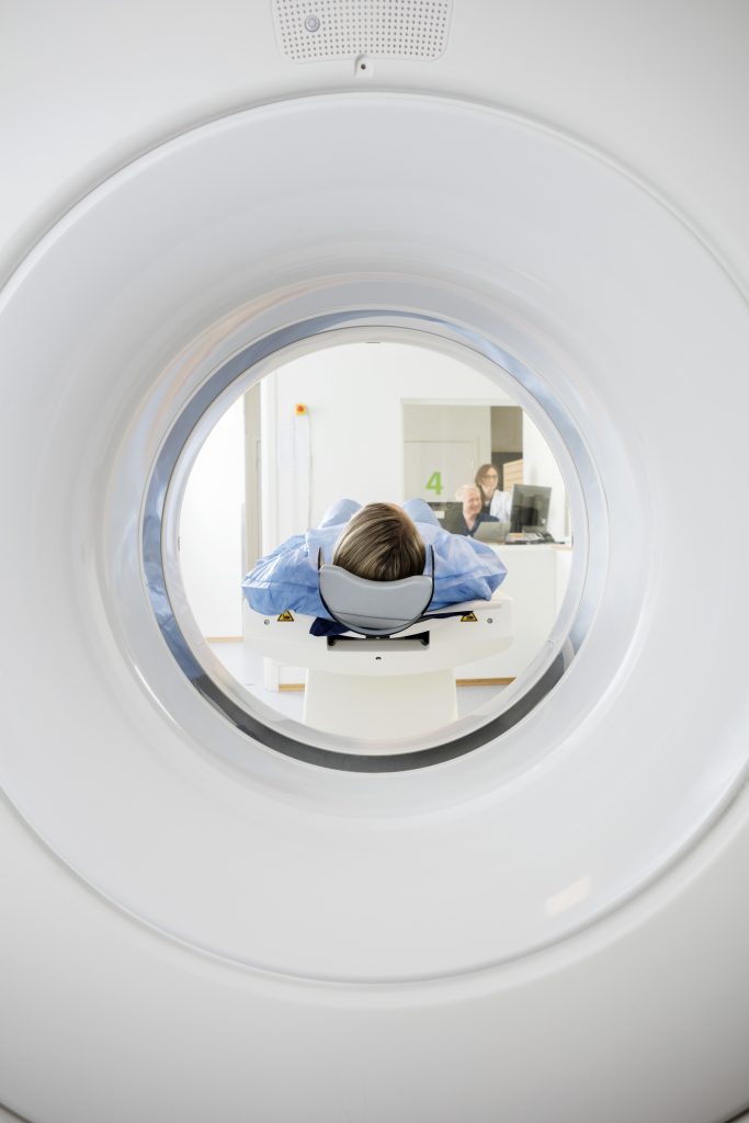
Cardiovascular computed tomography, or cardiac CT, is a noninvasive imaging test used to diagnose problems with the heart or peripheral arteries.
What is Computed Tomography?
Computed tomography (CT) is a type of imaging test that uses a combination of X-rays and computers to produce pictures of the vasculature. Today’s CT scanners are fast and can provide very detailed images of the arteries and veins.
When is Cardiac CT Used?
A cardiac CT can give your doctor more information on:
- The structure of your heart
- How well the heart pumps blood
- Scarring of the heart muscle caused by heart attacks
- Your risk of heart attack
- How much plaque buildup there is in the coronary arteries
- Possible abnormalities in the large blood vessels of the heart
The specific capabilities of a cardiac CT depend on whether or not a special contrast dye containing iodine is used. The contrast dye travels through the veins and shows up on the CT image.
Cardiac CT With Contrast
When a contrast dye is used, a cardiovascular CT scan can:
- Show blockages in the arteries of the heart
- Check for congenital heart defects
- Check how the ventricles of the heart are working
Cardiac CT Without Contrast
A cardiovascular CT without a contrast dye can:
- Measure the amount of calcium in the heart’s arteries (called a calcium score)
Vascular CT Angiography
Creates detailed images of the peripheral arteries:
- Carotid
- Abdominal Aorta
- With and without runoff
- Pulmonary
- Renal
- Thoracic Aorta
Risks of Cardiovascular CT
A cardiac CT scan is painless and is noninvasive unless a contrast dye is used. The only risks associated with cardiac computed tomography scans are associated with the low doses of radiation used to get the images. CT scans expose you to a small amount of X-ray radiation, so pregnant women and people without signs of heart problems should not take this test.
Preparing for Cardiac CT
Your doctor will give you instructions on how to prepare for the cardiac computed tomography scan. If contrast dye is going to be used, you shouldn’t eat for at least four to six hours before the test. If you aren’t given a contrast dye, then you should not eat for at least two hours before the test. Again, your doctor should give you specific instructions on what you need to do before the test. If you have any questions, be sure to ask.
What Happens During the Test?
Cardiovascular CT scans are performed at outpatient clinics or in hospitals. The test itself does not take long.
- If contrast dye is being used, it will be injected into an arm vein with an IV.
- During the test, you lie down on a table in the CT scanner.
- Electrodes are attached to your chest to monitor your heart. It is also connected to the CT scanner to create a clear picture of the heart.
- The table will slowly move inside the machine. The machine will arch around you, but it will not touch you and the scan is painless.
- The technologist administering the test will watch you closely through a window.
- You can talk to the technologist through a two-way intercom.
- The technologist will have you hold your breath for short periods of time.
- The entire scan should take about 5-10 minutes.
The physicians at Clearwater Cardiovascular Consultants provide a full range of outpatient cardiovascular services, including cardiac CT. We offer patients comprehensive care that is backed by experience, expertise, and the latest technology. Call Clearwater Cardiovascular Consultants at 727-445-1911 to make an appointment, or request an appointment online.
